The universal messenger in cellular biology

In the complex world of molecular biology, few ions carry as much weight as calcium. It is often referred to as a ‘universal messenger’ because it regulates an almost dizzying array of cellular processes, from the initial moments of fertilisation to the final stages of programmed cell death. Because calcium levels inside a cell are so tightly controlled and so reactive to external stimuli, measuring these changes has become a fundamental practice in laboratory research. This is where the calcium assay comes into play, serving as a vital window into the inner workings of living cells.
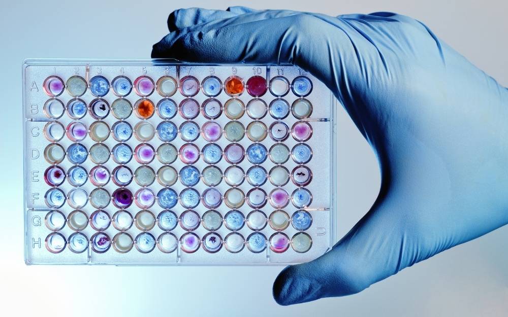
When we talk about a calcium assay, we are essentially describing a method used to monitor the concentration of calcium ions (Ca2+) within a biological sample. Most often, this involves looking at the cytoplasm of living cells. Because the resting concentration of calcium in the cytosol is kept incredibly low compared to the extracellular environment, even a small influx of ions creates a massive relative change. This signal is what researchers are looking for, as it usually indicates that a cell has responded to a drug, a hormone, or an electrical impulse.
How we actually see what the calcium is doing
Since calcium ions are invisible to the naked eye and do not naturally emit a signal that we can detect with standard microscopes, scientists have had to get creative. The most common way to perform a calcium assay is by using fluorescent indicators. These are specialised molecules designed to enter the cell and change their light-emitting properties when they bind to calcium ions.
The process typically follows a specific sequence of events:
- Dye Loading: Cells are incubated with a membrane-permeant version of a fluorescent dye. Once inside the cell, enzymes called esterases cleave the molecule, trapping it within the cytoplasm.
- Stimulation: The researcher introduces a stimulus, such as a new drug candidate or a neurotransmitter, to see if it triggers a calcium response.
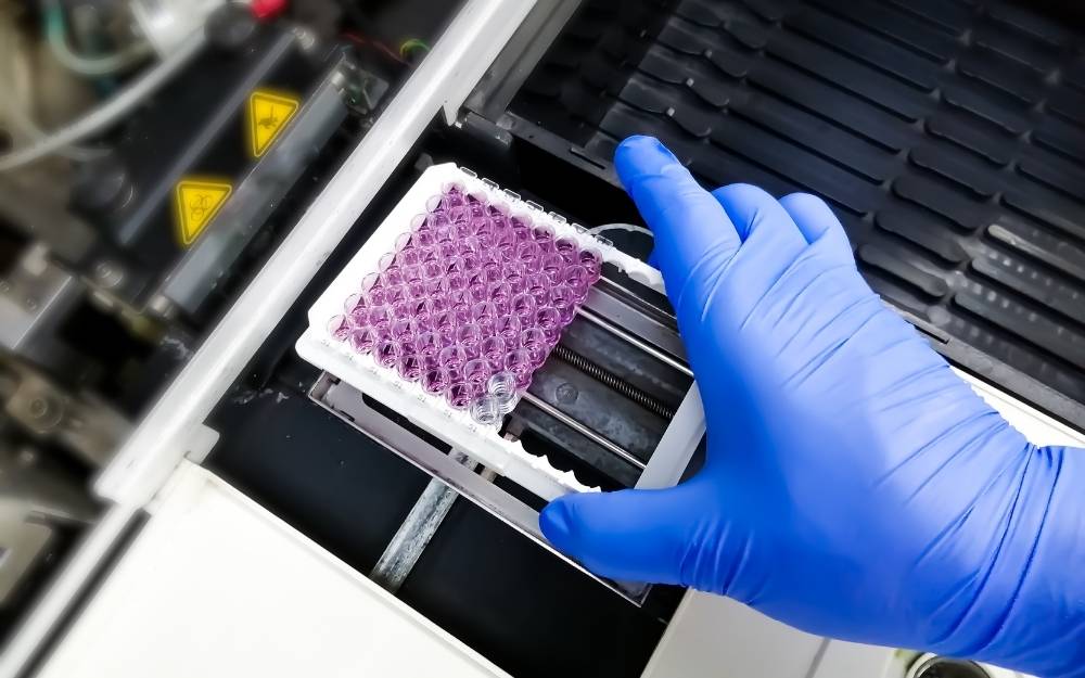
- Detection: A plate reader or a fluorescence microscope measures the change in light intensity or colour, which correlates directly to the amount of calcium present.
There are two main categories of these fluorescent indicators: ratiometric and single-wavelength dyes. Ratiometric dyes, like Fura-2, are particularly clever because they shift their peak excitation or emission wavelength when they bind to calcium. By taking a ratio of the signals at two different wavelengths, researchers can cancel out variables like uneven dye loading or cell thickness, leading to much more accurate data. Single-wavelength dyes, such as Fluo-4, are simpler to use and provide a much brighter signal, making them ideal for high-throughput screening where speed is more important than absolute quantification.
Why the drug discovery industry is obsessed with calcium
If you look at the portfolio of any major pharmaceutical company, a significant portion of their targets will involve G protein-coupled receptors (GPCRs) or ion channels. These are the ‘gatekeepers’ of the cell membrane, and a huge number of them use calcium as their primary downstream signalling mechanism. This is why the calcium assay has become the workhorse of the drug discovery industry.
When a new chemical compound is being tested, researchers need to know if it actually ‘turns on’ or ‘turns off’ the target receptor. By monitoring the calcium flux in real-time, they can see exactly how the cell responds. If a compound is designed to treat hypertension, for example, researchers might look for its ability to block calcium entry into vascular smooth muscle cells, which would prevent them from contracting too forcefully.
Beyond simple ‘yes or no’ answers, these assays allow scientists to determine:
- Potency: How much of the drug is needed to elicit a half-maximal response (EC50).
- Efficacy: The maximum response the drug can produce compared to a known standard.
- Kinetics: How quickly the calcium levels rise and how long it takes for them to return to baseline.
The critical role of calcium in cardiac research
One of the most specialised applications of this technology is in the study of the heart. In cardiac myocytes (heart muscle cells), calcium is the literal link between electrical excitation and physical contraction. This process, known as excitation-contraction coupling, must be perfectly timed for the heart to pump blood efficiently. Any disruption in this calcium cycle can lead to arrhythmias or heart failure.
Modern cardiac safety testing relies heavily on the calcium assay to ensure that new drugs don’t have unintended side effects on the heart. By using human induced pluripotent stem cell-derived cardiomyocytes (hiPSC-CMs), researchers can create a ‘heart in a dish’ and monitor its calcium handling. This provides a much more accurate representation of human biology than older methods that relied solely on animal models or simple cell lines. Observing the calcium transient—the rise and fall of calcium with every beat—allows scientists to spot subtle irregularities that might indicate a risk of pro-arrhythmia long before a drug ever reaches clinical trials.
Technical hurdles and how to overcome them
While the concept of measuring calcium seems straightforward, the practical execution requires a lot of finesse. One of the biggest challenges is ‘background noise.’ Many chemical compounds used in drug libraries are naturally fluorescent, which can interfere with the signal from the calcium dye. To combat this, researchers often use ‘masking dyes’ that stay outside the cell and quench any fluorescence that isn’t coming from the cytoplasm.
Another issue is dye leakage. Some cell types have active transporters that try to pump the fluorescent dye back out of the cell as soon as it is loaded. To prevent this, inhibitors like probenecid are added to the assay buffer to keep the dye where it belongs. Furthermore, the temperature must be strictly controlled; many calcium-handling proteins are highly sensitive to thermal changes, and performing an assay at room temperature when it should be at 37°C can lead to misleading results.
The move towards bioluminescence
While fluorescence is the dominant method, bioluminescence is a fascinating alternative that is gaining traction for specific uses. This involves using proteins like Aequorin, which is derived from jellyfish. Unlike fluorescent dyes, Aequorin doesn’t need an external light source to work; it emits light naturally when it binds to calcium. The main advantage here is the complete lack of background autofluorescence, resulting in a incredibly high signal-to-noise ratio. However, it requires the cells to be genetically engineered to express the protein, which adds a layer of complexity to the experimental setup.
As we look toward the future, the integration of automation and high-resolution imaging is making these assays faster and more detailed than ever. We are moving away from simply measuring a ‘bulk’ signal from a whole well of cells and moving toward single-cell analysis. This allows researchers to see how individual cells within a population behave differently, which is crucial for understanding complex diseases like cancer or neurodegeneration where cell heterogeneity is a major factor. The ability to organise and interpret this massive amount of data is the next great challenge in the field, requiring sophisticated software and machine learning to recognise patterns in the calcium oscillations that the human eye might miss.